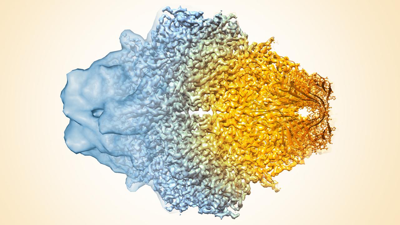
Inside every human cell is a microscopic world that scientists are still learning to see. A newly reported laser-powered improvement to electron microscopy may help reveal some of its smallest working parts.
The invisible world that controls our lives
Every second, countless molecular events take place inside the human body. Proteins fold, move, bind to partners and help cells respond to their surroundings. Yet many of these structures are so small that even sophisticated microscopes struggle to produce clear pictures of them. Scientists can identify genes and measure biochemical reactions, but understanding exactly how a molecular machine works often requires seeing its three-dimensional shape. A newly reported advance in electron microscopy may make that task easier. It is not a camera that can look through an entire living person, nor does it instantly diagnose disease. Instead, it improves one of the most demanding methods used to examine biological molecules.
What researchers announced in October 2026
Research from the University of California, Berkeley, reported on October 22, 2026, describes a laser phase plate designed to improve contrast in cryo-electron microscopy, commonly called cryo-EM. The device uses carefully controlled light to change the phase of electrons passing through an electron microscope. This approach is intended to make weakly visible molecular structures stand out more clearly. The important distinction is between a promising imaging advance and a routine, clinically proven tool: scientists must still establish performance across different specimens and research settings.
Why ordinary light microscopes are not enough
A familiar classroom microscope shines visible light through or onto a specimen. Its resolution is fundamentally limited by the wavelength of that light and the optical system. Human cells may be easy to see, but many individual proteins are only a few nanometres across. Electron microscopes use beams of electrons, whose wave properties allow much finer structural detail. However, achieving theoretical resolution is not the same as obtaining a clear image. Biological samples can be fragile, and tiny particles produce weak contrast against the surrounding material.
What cryo-electron microscopy actually does
In cryo-EM, researchers rapidly freeze a thin sample so that molecules are preserved in a glass-like layer of ice. The sample is then examined with an electron beam at very low temperature. Many images of molecules in different orientations can be combined computationally to reconstruct their structures. This method has helped scientists study ribosomes, viruses, membrane proteins and other molecular systems. It also has limitations: specimens can move, electrons can damage samples, images may be noisy, and small proteins can be particularly difficult to reconstruct.
The central problem: seeing objects that barely change the image
Some biological molecules do not strongly absorb or scatter enough electrons to generate a distinct picture. They can alter the phase of an electron wave without producing an obvious intensity difference at the detector. This is analogous to trying to see a nearly transparent object against a nearly transparent background. Phase-contrast methods translate otherwise subtle wave changes into visible differences. The concept transformed light microscopy decades ago, but implementing a powerful, stable phase plate inside an electron microscope poses major technical challenges.
How a laser phase plate changes the picture
The Berkeley approach uses an intense, carefully controlled laser field to influence the phase of electrons. In principle, that makes details from weakly scattering biological molecules more visible without relying only on conventional defocus-based contrast. Researchers must precisely align the laser and electron beams and manage the demanding optical conditions inside the instrument. This is not a laser that illuminates the cell directly like a flashlight; it is part of the electron-optical system. Improved contrast may reduce some of the difficulty of detecting and aligning very small particles for three-dimensional reconstruction.
Why tiny proteins matter so much
Proteins are involved in almost every major process in biology. Some carry chemical signals, some transport molecules, some build cellular structures, and others repair damage. A protein's function depends partly on its shape and how that shape changes. Researchers investigating a disease often need to understand how a protein interacts with another molecule or how a mutation alters its behaviour. Better structural imaging can support those investigations, especially where current techniques produce incomplete or uncertain models. But a clearer structure alone does not explain every aspect of a disease.
Could this accelerate drug discovery?
Many medicines work by binding to proteins or other molecular targets. Structural information can help scientists identify binding pockets, compare candidate compounds and explain why one molecule behaves differently from another. If new microscopy tools make difficult proteins easier to image, they could expand the range of targets that researchers can study. Drug discovery still requires extensive experiments on safety, delivery, biological activity and effectiveness. A new microscope is a research instrument, not a shortcut past clinical trials or regulatory review.
The connection to cancer and infectious disease research
Cancer research frequently examines proteins that regulate cell division, DNA repair and communication between cells. Infectious disease research studies viral proteins and the cellular machinery pathogens exploit. Cryo-EM already contributes to these fields by revealing structural details that other methods may miss. Higher contrast could help scientists tackle smaller or more flexible components of these systems. Whether it leads to specific new medicines depends on future research; no particular cure should be inferred from the announcement.
The engineering challenges still ahead
Breakthrough announcements often describe the capability of a prototype or a research system. Widespread adoption depends on reliability, cost, ease of use, maintenance and compatibility with existing microscopes. Scientists will need to compare results on representative biological specimens, quantify improvements and test reproducibility. Laser systems add optical and alignment complexity. The key question is not merely whether a better image can be obtained in a demonstration, but whether researchers can obtain better data consistently and efficiently.
Why this discovery matters for India
India has growing research activity in structural biology, biotechnology, pharmaceuticals and advanced instrumentation. Improvements in microscopy could benefit scientists studying infectious diseases, enzymes, agriculture and molecular medicine. Yet sophisticated instruments require more than purchasing hardware: they need trained specialists, stable facilities, sample preparation expertise and computational infrastructure. Collaborative facilities, open training and access to high-quality research equipment may matter as much as the instrument itself. These are potential opportunities, not announced Indian deployment plans.
The broader lesson: science advances by improving what we can measure
Many scientific revolutions begin with better tools. Telescopes changed astronomy; sequencing technologies changed genetics; microscopy transformed cell biology. A new imaging technique can open questions that researchers previously could not investigate properly. The laser phase plate is exciting for the same reason: it may allow scientists to obtain structural evidence from molecules that have remained difficult to see. Its eventual importance will be measured through independent experiments and the discoveries those experiments enable.
What students should learn from this story
For students preparing for competitive examinations or pursuing science careers, this development connects wave optics, electromagnetic radiation, the wave nature of electrons, microscopy, proteins and biotechnology. It also illustrates how physics and biology interact: an optical innovation can become a tool for molecular medicine. A useful study exercise is to compare optical microscopy, conventional electron microscopy and cryo-EM, then explain why contrast and resolution are different concepts. The best scientific reading asks three questions: what was demonstrated, what is only anticipated, and what evidence is still missing.
Conclusion: a clearer view, not an instant cure
The October 2026 research offers a promising way to improve the visibility of the molecular structures that help cells function. Its appeal lies in the possibility of studying smaller and more challenging biological targets. It does not mean scientists can now see every molecule in a living cell, and it does not establish that new treatments will follow immediately. But if the technology proves robust, it could become an important addition to the instruments that drive modern biological discovery.
Frequently Asked Questions
What is a laser phase plate?
It is a device that uses a controlled laser field to change the phase of electrons, potentially improving image contrast in an electron microscope.
Is this microscope already diagnosing diseases?
No. The reported development concerns research imaging, not an approved diagnostic test.
What is cryo-EM?
Cryo-electron microscopy images rapidly frozen biological samples using an electron beam and computational reconstruction.
Can this technology cure cancer?
No. It may help researchers study some proteins relevant to disease, but treatments require separate development and clinical testing.
Why are smaller proteins difficult to image?
Small proteins often produce weak contrast, making it harder to identify their orientations and reconstruct reliable structures.
Sources and further reading
Editorial note: This article is an educational explanation of reported research. Future medical uses are possibilities, not established outcomes. The cover photograph is illustrative and does not depict the specific Berkeley device.
 History Geography General Science Economy